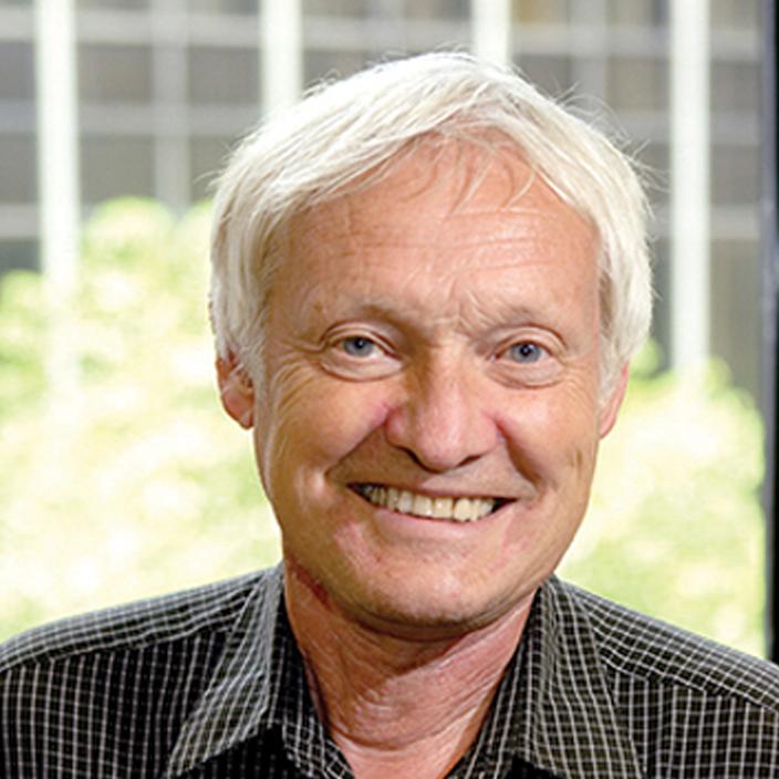
Much of biology comes down to studying the smaller pieces of the larger whole: the structure and workings of DNA, RNA, the synthesis and folding of the proteins through which all of life's workings are accomplished. But these intricate processes occur at a level of existence that requires sophisticated techniques to capture, study, and ultimately understand. Joachim Frank has dedicated his career to extending the vision of science to previously unseen layers and depths.
Ever since its invention, electron microscopy (EM) has been one of science's most powerful tools. Using a beam of electrons to probe matter at infinitesimal scales impossible with light microscopy, it has revolutionized the study of both the living and non-living universe. But it has its limitations, particularly in biology, where the radiation and hard vacuum needed for EM are anathema to living cells. Examining biological samples with EM generally means working with dead cells with a somewhat distorted structure unlike those in their native state. While dead cells are useful, their study doesn't allow in vivo visualization of living processes. Using the techniques of cryo-electron microscopy, Frank has overcome these difficulties and accomplished unprecedented feats of structural biology, including some of the most detailed images yet seen of the ribosome and its workings.
The ribosome, the complex molecular machine that translates messenger RNA into functional proteins, has been a central touchstone for most of Frank's work, both as a testing ground for the development of his microscopy and single-particle imaging techniques and as an object of study in its own right. Because the ribosome lacks the convenient crystallographic symmetry of other biological macromolecules, it has proven notoriously difficult to fully visualize at high resolution, but Frank made major strides in overcoming that problem. Devising multivariate statistical analysis techniques by which 2-D images from various angles (i.e., "single particles") could be combined and averaged to create 3-D images, Frank obtained views of the ribosome at far greater resolutions than had ever been obtained through conventional electron microscopy. He went on to develop the SPIDER software suite for the single-particle reconstruction of molecular structures, now used by researchers worldwide. In cryo-electron microscopy, a sample is examined after being frozen in vitreous (uncrystallized) ice, allowing biological macromolecules to be examined in their natural state without staining or other artifacts that can obscure structural detail. Frank used his image processing techniques in conjunction with cryo-EM to visualize the ribosome in action, showing protein synthesis as it happens. Perhaps his most notable achievement along these lines has been his discovery of the "ratcheting" motion that moves tRNA and mRNA through different parts of the ribosome during translocation. Rather than maintaining a static structure, the ribosome actually changes its conformational state as it performs its various functions. Frank continues his efforts to visually capture all of the ribosome's contortions as it synthesizes the proteins essential to life. His work not only influences the ways structural biology is done, but it also provides new views of the machinery of life at its most fundamental level.
Now a professor in the Department of Biochemistry and Molecular Biophysics and in the Department of Biological Sciences at Columbia University, and a Howard Hughes Medical Institute Investigator, Frank has published widely, producing more than 200 journal papers and six books. He is also a published author of poems and short stories, exploring the intricacies of the human heart with the same keen insight and perception that he brings to his scientific work.
Information as of April 2014

